
Optical Coherence Tomography Imaging
Highspeed, cross-sectional, label-free imaging modality, analogous to ultrasound, utilizing light instead of sound to create a gray-scale image from the echo time delay of infrared light reflected from the specimen.
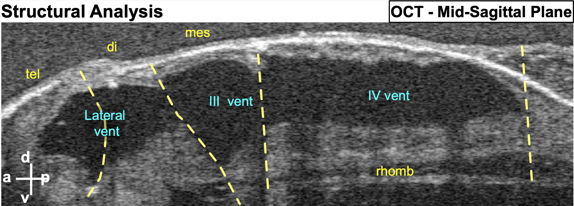
(a) Mid-sagittal view of the brain allowing simultaneous visualization of the four ventricles
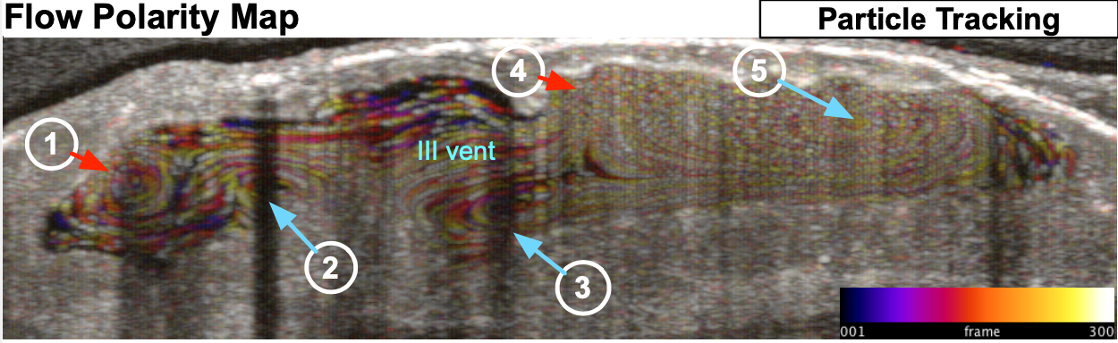
(c) Intrinsic Particle Tracking: Using ImageJ, we tracked particles using 300 frames of 2D images. Temporal color coding depicts their trajectory over time. Based on the trajectory map, 5 distinct flow fields were observed: FF1-FF5.
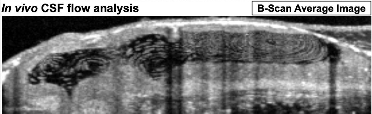
(b) B-scan image acquisition using OCT: Ventricles of a stage 45 tadpole harbor native free floating particles. Spatial position of the particles recorded over time to demonstrate discrete flow patterns within each ventricle
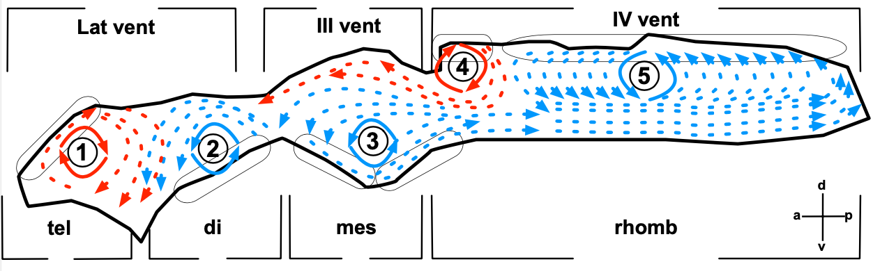
(d) Outline of the ventricular system and vector maps of the CSF flow fields. Based on the trajectory map and real time observation